

Copyrights: D. A. Omoboyowa, F. O. Afolabi, T. C. Aribigbola, 2018. License: This work is licensed under a Creative Commons Attribution 4.0 International License.
Abstract
Background: The anti-hyperglycemic potential of methanol stem bark extract of Anacardium occidentale (MSBEAO) was investigated using an alloxan-induced diabetic rat model. Alloxan administration induces the generation of free radicals which can affect antioxidant status resulting in the disruption of the β-cells of the pancreas. Therefore, this study examines the antioxidant potential of the plant extract and the ameliorating effect on the pancreas of alloxan-induced diabetic rats.
Methods: Diabetes was induced by intraperitoneal injection of 150 mg/kg body weight of alloxan monohydrate. MSBEAO, at a concentration of 100 or 200 mg/kg b.w. was orally administered to alloxan-induced diabetic rats and normal rats. The hypoglycemic effect, oral glucose tolerance test, and biochemical assay of alloxan-induced diabetic rats were assayed using standard procedures.
Results: Preliminary phytochemical screening of the extract revealed the presence of alkaloids, tannins, saponins, terpenoids, carbohydrates, and phenols at moderate concentrations. The lethality dose (LD50) of the plant extract was found to be equal to or less than 5000 mg/kg b.w. The hypoglycemic effect of the extract on the non-diabetic rats revealed a significant (p<0.05) decrease in the blood glucose concentration of animals administered with 1 g/kg b.w. of the extract, compared to normal control rats administered with normal saline. In the oral glucose tolerance test, the methanol extract exerted the highest response, similar to glibenclamide after 15 and 30 minutes of administration, compared to the control rats. The methanol extract yielded the highest blood glucose lowering effects after 9 days of treatment (p<0.05), compared to diabetic rats administered with normal saline and 0.3 mg/kg b.w. of glibenclamide. Administration of the extract at 200 mg/kg b.w. showed improved pancreas architecture and regeneration of the β-cells, compared with the pancreas of animals in the other groups.
Conclusion: The results of this study suggest that MSBEAO is a potentially effective agent for the management of diabetes which might result from the antioxidant-generating capacity of the stem bark.
Background
Diabetes mellitus (DM) is a type of metabolic disease in which there is a high blood glucose level over a prolonged period. It is a common endocrine disorder which is rapidly increasing in the human population all over the world 1. Diabetes often leads to complications such as peripheral neuropathy, retinopathy, coronary heart disease, and cataract 2. Persistent hyperglycemia causes increased generation of free radicals, especially reactive oxygen species, which may result from glucose auto-oxidation and protein glycosylation 3. Free radicals play an important role in the causation and complication of diabetes mellitus through the alterations in endogenous free radical scavenging defense mechanisms. While chemotherapy remains the major solution to diabetes control, the setbacks being encountered in present anti-diabetic therapies calls for more innovative treatment therapies that are effective, less toxic, less expensive, and with fewer side effects compared to synthetic drugs 4.
Cashew (Anacardium occidentale) is a well-known member of the Anacardiaceae family and is commonly found in Northeastern Brazil 5. The plant is commonly referred to as cashew in English, kashu in Hausa, and kaju in Yoruba. It has a thick and tortuous trunk with branches so winding that they frequently reach the ground 6. The cashew tree produces many resources and products. All parts of the plants, like leaves, fruits, and bark, have been traditionally used to relieve a variety of ailments 7. The kernel has been reported by Omoboyowa et al. 8 to possess anti-diarrheal effect with reversal of electrolyte imbalance in castor oil-induced diarrheal rats 5. However, this study is aimed at evaluating the possible anti-diabetic potential of methanol extract of Anacardium occidentale stem bark on the blood glucose profile of alloxan-induced diabetic and normal rats.
Methods
Plant Materials
The stem barks of A. occidentale were collected from trees within the Botanical Garden of Akanu Ibiam Federal Polytechnic, Unwana, Afikpo, Ebonyi State, Nigeria. The plant was authenticated by the Botany Department, University of Nigeria, Nsukka, Nigeria, where voucher specimens have been deposited (UNN/BOT/2016/058). The stem barks were shed dried and powdered for extraction. The air-dried powder was extracted using soxhlet extraction apparatus. One hundred and twenty grams (120 g) of the powder was extracted with 1000 ml of methanol for 48 hours. The extract obtained was evaporated under a vacuum evaporator. The yield of crude extract of A. occidentale stem bark was recorded to be 8.50 g (7.08 %). The extract was preserved in the refrigerator at 10oC during the experimental protocol. The methanolic extract of A. occidentale stem bark was prepared in normal saline for oral administration 9.
Chemical Reagents and Drugs
All the reagents used in this study were of analytical grade, and the diagnostic kits used for the estimation of serum catalase, superoxide dismutase (SOD), and glutathione (GSH) were obtained from Randox Diagnostic (England). Alloxan was purchased from Sigma-Aldrich (India) and standard glibenclamide was supplied by New Good Health Pharmacy (Nigeria).
Animals
Healthy male albino rats weighing 146 g and 196 g were procured from the Department of Veterinary Medicine, University of Nigeria Nsukka, Nigeria and maintained in standard animal cages at a temperature of 25±5 oC with 12-hour light and dark cycle. The animal experiments were conducted as per the AIFPU Scientific Research Committee in Nigeria. The experimental protocol was approved by the Institutional Animal Ethics Committee. During the animal experiments, animals were fed with standard pellet diet and free water ad libitum and allowed to acclimatize for seven days before they were randomly grouped for the experiment. Fifteen rats were used for the hypoglycemic study while twenty-five rats were grouped for the acute anti-hyperglycemic study.
Induction of Diabetes and Experimental Design
Diabetes was induced by intraperitoneal injection of alloxan monohydrate dissolved in normal saline at a dose of 150 mg/kg body weight in animals fasted overnight. After 48 hours, blood samples were collected from the tail vein and the blood glucose level was determined by a glucometer (AccuChek) 9. The animals showing blood glucose level higher than 200 mg/dL were considered as hyperglycemic and selected for the studies. The animals were divided into five groups containing five animals in each group (n = 5) as follows:
Group 1 – Non-diabetic control
Group 2 – Alloxan-induced diabetic rats administered with 0.3 ml of normal saline
Group 3 – Alloxan-induced diabetic rats treated with 0.3 mg/kg b.w. of Glibenclamide
Group 4 – Alloxan-induced diabetic rats treated with 100 mg/kg b.w. dose of MSBEAO;
Group 5 – Alloxan-induced diabetic rats treated with 200 mg/kg b.w. dose of MSBEAO.
Glibenclamide was used as the reference standard during the studies. After 72 hours of induction of diabetes, the plant extract suspended in normal saline was administered for 9 days using an oral feeding needle. Blood samples were collected on days 0, 3, 6, and 9 from the start of the study for measurement of blood glucose via the tail vein. Blood glucose levels were measured by glucometer. After 9 days, blood samples were collected through the retro-orbital puncture and analyzed for selected antioxidants. The pancreases were also collected for histopathological studies.
Phytochemical Test
The qualitative phytochemical screening of the methanol extract of A. occidentale stem bark was carried out using procedures outlined by Evans et al. 10.
Acute Toxicity and Lethality (LD50) Test
The acute toxicity and lethality of MSBEAO were determined using the method outlined by Omoboyowa et al. 9. The test was divided into two phases. In phase one, sixteen (16) randomly selected adult mice were divided into four groups, four per group (n = 4), and received 100, 300, 600 or 1000 mg/kg body weight of methanol extract. Signs of toxicity and possible death were monitored and recorded for a period of 24 hours. After the 24-hour observation, the doses for the second phase were determined based on the outcome of the first phase. Since there was no death, a fresh batch of animals was used following the same procedure in phase 1 but with higher doses (of 1900, 2600 or 5,000 mg/kg body weight) of extract. The animals were also observed for 24 hours for signs of toxicity and possible death. The LD50 was calculated as the geometric mean of the high non-lethal dose and lowest lethal dose 9.
Glucose Profile Study
Hypoglycemic Test in Non-Diabetic Rats
In this test, fifteen (15) non-diabetic rats were fasted overnight and divided evenly into three (3) groups. Group 1 rats were administered with 0.3 ml of normal saline, Group 2 rats were given 0.3 mg/kg body weight of glibenclamide, and Group 3 rats were given 1 g/mg body weight of MSBEOA. Blood samples were collected from the tail vein prior to and at 0, 1, 2, 3 and 5 hours after dosing, and used to estimate the glucose concentrations.
Oral Glucose Tolerance Test (OGTT) in Normal Rats
The procedure, dosage of extracts, normal saline and glibenclamide, and animal groupings in this study were as described in previous reports. In addition to the protocols, the rats were orally administered with glucose (2 g/kg body weight) 30 min after dosing, and blood samples were obtained via the tail vein at time 0 (prior to glucose dosing), at 15, 30, 45, 60, 90 or 120 min after glucose administration, to measure the glucose levels.
Acute Anti-Hyperglycemic Study
Glucose profile studies were conducted with non-diabetic rats and alloxan-induced diabetic rats as described by Atangwho et al. 11.
In this test, 4 groups of alloxan-induced diabetic rats were treated as follows: Group II received 0.3 ml of normal saline, Group III received 0.3 mg/kg body of Glibenclamide, Group IV received 100 mg/kg body weight of MSBEAO, Group V received 200 mg/kg MSBEAO, and Group 1 served as the normal control group. The extract, normal saline and glibenclamide were administered once per day during the 9-day period of the study. Fasting blood glucose (FBG) was measured on day 0 (baseline), and days 3, 6 and 9. At the end of the study, the animals were euthanized, and then the pancreas was removed and preserved for histology. The AccuCheck glucometer was used to measure blood glucose levels.
Biochemical Assay
The concentrations of glutathione, malondialdehyde, and vitamin C, and catalase and superoxide dismutase activities were determined spectrophotometrically according to the Randox assay kit.
Pancreas Histopathological Study
Dissected pancreas from control, diabetic and treated albino rats were fixed in 10% formaldehyde and processed, and used for histopathological analysis. Tissue processing was carried out using an autotechnicon and the prepared, 5 µm thick sections were morphologically evaluated by an independent histopathologist, according to the method described by Ezejiofor et al. 12.
Measurement of Body and Organ Weight
The body weight and weights of select organs (spleen, liver, kidney, and heart) were determined using high precision balance (HPB 2000-10 mg, China).
Statistical Analysis
Statistical analyses of all the results were performed by one-way analysis of variance, followed by post hoc multiple comparison tests using the SPSS (version 16) software. Results were expressed as mean ± standard error of mean (SEM). The statistical significance level was set at p < 0.05.
Results
Lethal Dose (LD50) Results
In the experiment, there was no lethality or behavioral changes in the three groups of the mice that received 10, 100, or 1000 mg/kg body weight of the MSBEAO at the end of the first experiment. Further dose increase to 1900, 2600 and 5000 mg/kg body weight of the extract did not induce death within 24 hours of administration. Together, these results showed that the extract was relatively safe at doses equal to or greater than 5000 mg/kg body weight.
Phytochemical Screening
The phytochemical study of A. occidentale stem bark revealed a high presence of tannins, saponins, terpenoids, carbohydrates, and phenols, a moderate presence of anthraquinones, but low presence of steroids, alkaloids, flavonoids, and reducing sugars, as indicated in Table 1.
| Phytochemical Compounds | Result |
| Steroids | + |
| AlkaloidsWagner’s test | ++ |
| Tannins | +++ |
| SaponinsFroth test | ++ |
| Flavonoids | + |
| Terpenoids | ++ |
| Reducing sugars | + |
| Phenols | ++ |
| Anthraquinones | ++ |
| CarbohydratesFehling’s test | ++++ |
Hypoglycemic Test in Non-Diabetic Animals
The effects of the methanol extract of A. occidentale stem bark and glibenclamide on the glucose levels of non-diabetic rats are shown in Figure 1. The diabetic animals administered with 0.3 mg/kg body weight of glibenclamide and 1 g/kg body weight of MSBEAO showed a significant (p<0.05) decrease in glucose levels throughout the experimental period as compared with normal control animals. The hypoglycemic effect of the standard drug (glibenclamide) was observed to be significantly (p<0.05) higher compared with that of the extract.

Oral Glucose Tolerance Test on Non-Diabetic Rats
The effects of MSBEAO and glibenclamide on oral glucose tolerance test of non-diabetic rats are shown in Figure 2. The measured FBG reached its peak value 15 minutes after oral administration of glucose. Animals administered with 2 g/kg body weight of glucose and 0.3 mg/kg body weight of glibenclamide had the most significant (p<0.05) reduction in FBG; the reduction was sustained throughout all the measured times compared to the glucose levels of the other treatment groups. The animals administered with 2 g/kg body weight of glucose and 1 g/kg body weight of MSBEAO showed a significant (p<0.05) increase in glucose levels after 30 min of treatment compared to glucose levels after 15 min of treatment. There was also a significant (p<0.05) reduction in glucose levels after 45, 60, 90 and 120 min, respectively, compared to glucose levels after 30 min.

Body Weight of Treated and Non-Treated Alloxan-Induced Diabetic Animals
The effects of MSBEAO and glibenclamide on body weight of alloxan-induced diabetic rats are shown in Figure 3. Diabetic animals treated with 100 mg/kg body weight of MSBEAO showed a significant (p<0.05) increase in body weight on days 0, 3, 6 and 9, respectively, compared to animals treated with 0.3 mg/kg body weight of glibenclamide. Animals induced and treated with 200 mg/kg body weight of MSBEAO showed a significant (p<0.05) reduction in body weight on days 0, 3 and 6, respectively, compared to animals treated with 100 mg/kg body weight of MSBEAO.

Weight of Selected Organs of Treated and Non-Treated Alloxan-Induced Diabetic Rats
The effects of MSBEAO and glibenclamide on weight of select organs (spleen, liver, kidney, and heart) in alloxan-induced diabetic rats are shown in Figure 4. Diabetic-induced animals treated with 0.3 mg/kg body weight of glibenclamide showed a significant (p<0.05) reduction in the weight of spleen, liver, kidney and heart, respectively, compared to animals treated with 0.3 ml of normal saline. Animals induced and treated with 100 mg/kg body weight of the extract showed a significant (p<0.05) increase in the weight of spleen, liver, kidney and heart, respectively, compared to animals treated with 0.3 mg/kg body weight of glibenclamide. Animals induced and treated with 200 mg/kg body weight of the extract showed a significant (p<0.05) reduction in the weight of spleen, kidney and heart, respectively, compared to weight of animals treated with 100 mg/kg body weight of the extract. There was also a significant (p<0.05) increase in the weight of liver, compared to that of animals treated with 100 mg/kg body weight of MSBEAO.

Blood Glucose Levels of Treated and Non Treated Alloxan-induced Diabetic Animals
The effects of different dosages of MSBEAO on blood glucose levels in diabetic rats are illustrated in Figure 5. The animals treated with MSBEAO at 100 and 200 mg/kg b. w. showed significant (p<0.05) hypoglycemic activity on the 9th day compared with the diabetic control group. While the animals that received 100 mg/kg b. w. of the extract also showed non-significant (p>0.05) reduction in blood glucose concentration compared with the diabetic animals treated with 200 mg/kg b. w. of the extract. The animals that received glibenclamide at 0.3 mg/kg significantly (p<0.05) reduced the blood glucose level on the 9th day of the study compared with the diabetic control animals.

Effect of Methanol Extract of A. occidentale Stem Bark on Antioxidant Parameters of Alloxan-Induced Diabetic Rats
The alloxan-induced diabetic rats treated with 0.3 mg/kg body weight of glibenclamide and 100 mg/kg body weight of MSBEAO showed a significant (p<0.05) reduction in serum malondialdehyde concentration compared to the diabetic-induced animals administered with 0.3 ml of normal saline. The diabetic animals treated with 0.3 mg/kg body weight of glibenclamide and 100 mg/kg body weight of the extract showed a significant (p<0.05) reduction in vitamin C concentration and catalase activity compared to the alloxan-induced animals administered with 0.3 ml of normal saline. The glutathione concentration in the diabetic rats treated with 200 mg/kg body weight of the extract was significantly (p<0.05) higher compared to the diabetic animals treated with 100 mg/kg body weight of MSBEAO (Table 2).
| Treatments | Malondialdehyde (mg/ml) | Superoxide dismutase (µl) | Glutathione (mg/dl) | Vitamin C (mg/dl) | Catalase (µl) |
| NR | 2.80 ± 0.20 | 1.13 ± 0.002 | 4.20 ± 0.10 | 2.26 ± 0.05 | 6.45 ± 0.04 |
| AIRANS | 6.55 ± 0.25* | 0.91 ± 0.01* | 3.85 ± 0.15 | 2.28 ± 0.03 | 10.02 ± 0.10* |
| AIRAG | 4.80 ± 0.10* | 1.11 ± 0.01 | 5.65 ± 0.05* | 1.97 ± 0.13* | 5.57 ± 0.20* |
| AIRA100AO | 5.70 ± 0.20* | 1.06 ± 0.007* | 2.50 ± 0.50* | 1.81 ± 0.04* | 7.00 ± 0.08 |
| AIRA200AO | 5.05 ± 0.25* | 1.09 ± 0.008* | 4.05 ± 0.15 | 1.94 ± 0.04* | 12.85 ± 0.16 |
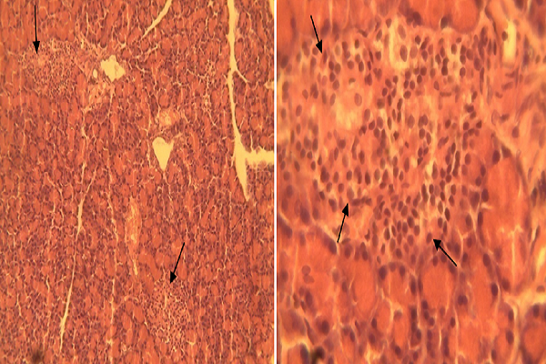
Histopathology of the Pancreas
The prepared slides were examined with a Motic™ compound light microscope using 4X, 10X and 40X objective lenses. The photomicrographs were taken using a Motic™ 9.0 megapixels microscope camera at 100X and 400X magnifications.


Discussion
Diabetes mellitus is a metabolic disease associated with impaired glucose metabolism which adversely alters the intermediary metabolism of lipids and carbohydrate. It is a chronic metabolic disorder of carbohydrate, fat and protein metabolism, characterized by elevation of both fasting and postprandial blood glucose levels 13. In the present study, the anti-hyperglycemic activity of the methanol extract of A. occidentale stem bark was evaluated in alloxan-induced diabetic and normal rats, using fasting blood glucose test. The results showed that serum glucose concentration in diabetic rats treated with 100 mg/kg and 200 mg/kg of extract was reduced significantly (p<0.05), and the blood glucose lowering effects of the extract were traceable to its constituents.
While saponins are known to have anti-hyperglycemic effect, flavonoids have been found to be an active principle in herbal medicine and are known to be powerful antioxidants that may protect organs against toxicity and potential damage due to agents such as alloxan 14. The induced weight loss observed in diabetic untreated rats, as a result of alloxan injection and compared to treated rats, mimic the common weight loss usually diagnosed in clinical diabetic patients. During the 9 days of treatment, continuous reduction in body weight of alloxan-induced diabetic rats was observed, whereas a significant (p<0.05) gain in body weight was observed in A. occidentale-treated rats. The treatment with A. occidentale extract ameliorated the loss in body weight and restored this level towards normal.
The potential of A. occidentale extract to correct the body weight might result from its anti-hyperglycemic ability by increasing the rate of glucose metabolism. Insufficient insulin secretion prevents the body from obtaining glucose from the blood into the body’s cells to be metabolized as energy. Therefore, the body starts to mobilize fat and muscle for energy, causing a reduction in overall body weight. The ability of A. occidentale extract to reduce hyperglycemia and protect against muscle wasting might increase the availability of glucose for energy production, thereby restoring body weight.
The present study examines the antioxidant potential of methanol stem bark extract of A. occidental in alloxan-induced diabetes rats. The results showed a high level of antioxidant potential which may play an important role in the management of disorders involving oxidative stress. Plants containing natural antioxidants, such as tannins, flavonoids, vitamin C and vitamin E, can preserve β–cell function and prevent diabetes-induced formation of reactive oxygen species, leading to inhibition of lipid peroxidation 149.
The anti-hyperglycemic properties of MSBEAO may be possible due to insulin-released stimulating effects and uptake of peripheral glucose, which in turn reverse alloxan-induced hyperglycemia. However, islet lesions and destruction of β-cells were evident in the untreated hyperglycemic group. Treatment with MSBEAO results in the proliferation of β-cells and reversal of islet lesions. This results in the upregulation of insulin secretion, inducing a reduction in hyperglycemia as observed in treated animals. The β-cells, which are the insulin- secreting cells, make up over 80% of the total number of cells in the pancreatic islets.
Alloxan causes necrosis of the β-cells of the pancreatic islets. This is the reason why there is a decrease in the number and the size of the pancreatic islets. In this study, the observations of Groups 1 (untreated control), 2 (alloxan only) and 3 (alloxan and standard drug) were as expected. However, the observations in the treated groups were unexpected. First, the low dose groups showed decreased size of the hypercellular islets, while the high dose groups showed relatively normal sized pancreatic islets. In the literature, β-cells are not known to regenerate in adult subjects but the findings in the high dose groups suggest that regeneration of β-cells may have occurred.
Conclusions
A. occidentale stem bark extract possessed anti-hyperglycemic activity like the standard drug used in this study. This was based on its prominent reduction of blood glucose levels in the animals treated with different doses compared with the untreated diabetic animals. The observed anti-hyperglycemic potential of the plant extract may be attributed to the presence of bioactive compounds and antioxidant-generating capacity. However, the results of this study justify the traditional use of the plant in the treatment of diabetes.
Open Access
This article is distributed under the terms of the Creative Commons Attribution License (CCBY4.0) which permits any use, distribution, and reproduction in any medium, provided the original author(s) and the source are credited.
List of abbreviations
DM: Diabetes mellitus; FBG: Fasting blood glucose; GSH: Glutathione; LD50: Lethal dose 50; MSBEAO: Methanol stem bark extract of Anacardium occidentale; SOD: Superoxide dismutase; SPSS: Statistical package for social sciences
Ethics approval and consent to participate
The animal experiments were conducted as per the AIFPU Scientific Research Committee in Nigeria.
Competing interests
The authors declare that they have no conflicts of interest.
Authors' contributions
This work was carried out in collaboration between all authors. Authors DAO, FOA and TCA designed the study, wrote the protocol and supervised the work. Authors DAO and TCA carried out all laboratories work. Author DAO performed the statistical analysis. Authors DAO and FOA managed the analyses of the study. Authors DAO and FOA wrote the first draft of the manuscript. Authors DAO and TCA managed the literature searches and edited the manuscript. All authors read and approved the final manuscript.
Acknowledgments
The authors are grateful to Mr. Eguonu M. O. and Dr. Ezeasor K. C. of the Department of Veterinary Pathology and Microbiology, Faculty of Veterinary Medicine, University of Nigeria, Nsukka for the preparation and interpretation of the pancreas histopathology.
References
-
Andrade
M.,
Helnrich
N..
The Rising Global Burden of Diabetes and its Complication. European Journal of Cardiovascular Prevention and Rehabilitation.
2005;
17
:
3-8
.
-
Matsuda
H.,
Murakami
T.,
Yashiro
K.,
Yamahara
J.,
Yoshikawa
M..
Antidiabetic principles of natural medicines. IV. Aldose reductase and qlpha-glucosidase inhibitors from the roots of Salacia oblonga Wall. (Celastraceae): structure of a new friedelane-type triterpene, kotalagenin 16-acetate. Chemical & Pharmaceutical Bulletin.
1999;
47
:
1725-9
.
-
Moussa
S. A..
Oxidative stress in Diabetes mellitus. Romanian Journal of Biophysics.
2008;
18
:
225-35
.
-
Iqbal
N..
The burden of type 2 diabetes: strategies to prevent or delay onset. 2007;
3
:
511-20
.
-
Omoboywa
D. A.,
Nwodo
O. F.,
Joshua
P. E.,
Akalonu
C. X..
Effect of chloroform-ethanol extracts of cashew (Anacadium occidentale) kernel on electrolyte imbalance in castor oil induced diarrhea rats. International Journal of Biochemistry Research & Review.
2015;
8
:
1-6
.
-
Adesokan
A.,
Oyewole
I.,
Turay
B..
Kidney and liver function Parameters in Alloxan induced Diabetic Rat Treated with Aloe Barbadenesis juice Extract. 2009;
1
:
33-7
.
-
Dare
S. S.,
Hamman
O.,
Musa
A..
Effects of Aqueous Extract of Anacardium occidentale (cashew) leaf on pregnancy outcome of wister Rat. International. Journal of Animal and Veterinary Advances.
2011;
3
:
77-82
.
-
Omoboyowa
D. A.,
Nwodo
O. F.,
Joshua
P. E..
Anti-diarrhoeal activity of chloroform-ethanol extracts of cashew (Anacadium occidentale) kernel. Journal of Natural Products.
2013;
6
:
109-17
.
-
Omoboyowa
D. A.,
Igara
E. C.,
Otuchristian
G.,
Olugu
K. D..
Antidiabetic activity of methanolic extract of seed cotyledon of Chrysophyllum albidum in alloxan-induced diabetic rats. Biokemistri.
2016;
28
:
88-95
.
-
Evans
William Charles.
2009.
Google Scholar -
Atangwho
I. J.,
Egbung
G. E.,
Ahmad
M.,
Yam
M. F.,
Asmawi
M. Z..
Antioxidant versus anti-diabetic properties of leaves from Vernonia amygdalina Del. growing in Malaysia. Food Chemistry.
2013;
141
:
3428-34
.
-
Ezejiofor
A. N.,
Okorie
A.,
Orisakwe
O. E..
Hypoglycaemic and tissue-protective effects of the aqueous extract of persea americana seeds on alloxan-induced albino rats. 2013;
20
:
31-9
.
-
Hussain
M. A.,
Theise
N. D..
Stem-cell therapy for diabetes mellitus. Lancet.
2004;
364
:
203-5
.
-
Hunt
J. V.,
Smith
C. C.,
Wolff
S. P..
Autoxidative glycosylation and possible involvement of peroxides and free radicals in LDL modification by glucose. Diabetes.
1990;
39
:
1420-4
.
Comments
Downloads
Article Details
Volume & Issue : Vol 5 No 7 (2018)
Page No.: 2440-2454
Published on: 2018-07-27
Citations
Copyrights & License
This work is licensed under a Creative Commons Attribution 4.0 International License.
Search Panel
Pubmed
Google Scholar
Pubmed
Google Scholar
Pubmed
Search for this article in:
Google Scholar
Researchgate
- HTML viewed - 4600 times
- Download PDF downloaded - 860 times
- View Article downloaded - 0 times
